
Dental X-Rays in Brentwood
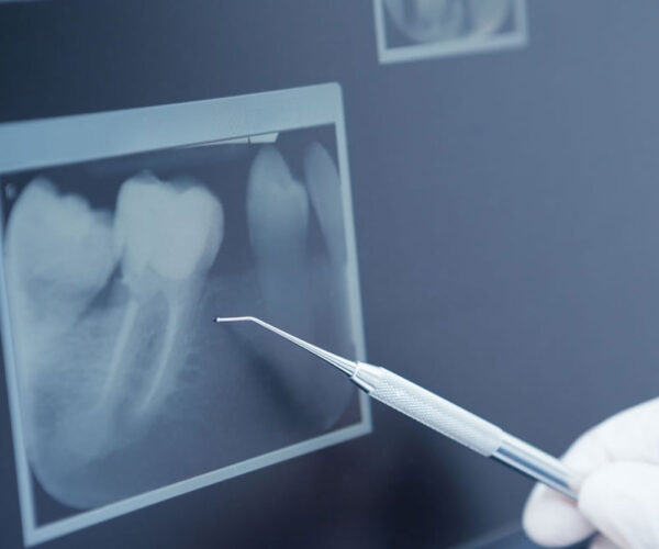
X-rays, also known as radiographs, are an essential part of any dental care treatment plan. They are diagnostic, but they can also be preventative, by helping a dentist diagnose potential oral care issues in a patient’s mouth before they become a major problem. An x-ray is a type of energy that passes through soft tissues and is absorbed by dense tissue. Teeth and bone are very dense, so they absorb X-rays, while X-rays pass more easily through gums and cheeks.
X-rays are divided into two main categories, intraoral and extraoral. Intraoral is an X-ray that is taken inside the mouth. An extraoral X-ray is taken outside of the mouth.
Intraoral X-rays are the most common type of radiograph taken in dentistry. They give a high level of detail of the tooth, bone and supporting tissues of the mouth. These X-rays allow dentists to:
- Find cavities
- Look at the tooth roots
- Check the health of the bony area around the tooth
- Determine if periodontal disease is an oral care issue
- See the status of developing teeth
- Otherwise, monitor good tooth health through prevention

Most popular and trending Dental X-Ray Questions
Most people with healthy teeth and gums should have dental X-rays taken once every six to 18 months. But if you have gum disease, recurring decay or other time-sensitive oral health issues, you may need more frequent X-rays.
There is no number that is definitely safe, just as there is no number that is definitely dangerous. Every x-ray can involve some tiny risk. If the x-ray is needed to find out about a medical problem, then that small risk is certainly worth taking.
An X-ray won’t show subtle bone injuries, soft tissue injuries or inflammation. However, even if your doctor suspects a soft tissue injury like a tendon tear, an X-ray might be ordered to rule out a fracture.